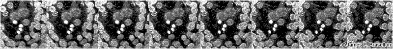
Incipient double fertilization in an intact ovule in a cross between Solanum stoloniferum (ovulate parent) and S. phureja (pollen donor), showing one sperm nucleus near the two polar nuclei and the other sperm nucleus near the egg nucleus. The other two brightly fluorescent objects are the x-bodies, one the remnants of the vegetative (tube) nucleus and the other the nucleus of the degenerated synergid.
These 8 confocal scanning laser microscopy images are presented so that every set of two side-by-side images forms a stereo pair with 7 degrees separation for viewing with a standard stereo viewer or map-reading device, or by relaxing and crossing your eyes until the right and left images of each pair fuse into one.
Reference: Braselton, J. P., M. J. Wilkinson, & S. A. Clulow. 1996. Feulgen staining of intact plant tissues for confocal microscopy. Biotech. & Histochem. 71: 84-87.