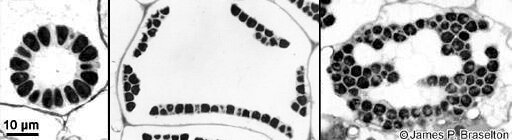
Light micrographs of sporeballs (sporosori aka cystosori) of Sorosphaera veronicae, Membranosorus heterantherae, and Spongospora subterranea. The material was fixed in glutaraldehyde and osmium tetroxide, embedded in epoxy plastic, and sectioned approximately 0.5 μm in thickness before viewing with the oil-immersion, 100X objective of the light microscope.
Reference: Bulman, S. and J. P. Braselton. 2014. Rhizaria: Phytomyxea. Pp. 99-112. In: McLaughlin, D. J. and J. W. Spatafora, eds. The Mycota VII, 2nd ed., Part A, Systematics and Evolution, Springer-Verlag, Berlin-Heidelberg.